Home

Ödland Spenden Stengel irma diabetic retinopathy Symposium sehen Entfernung

JCDR - Dyslipidemia, Lipid profile, Retinopathy, Type 2 diabetes

Diabetic retinopathy

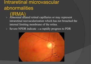
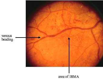
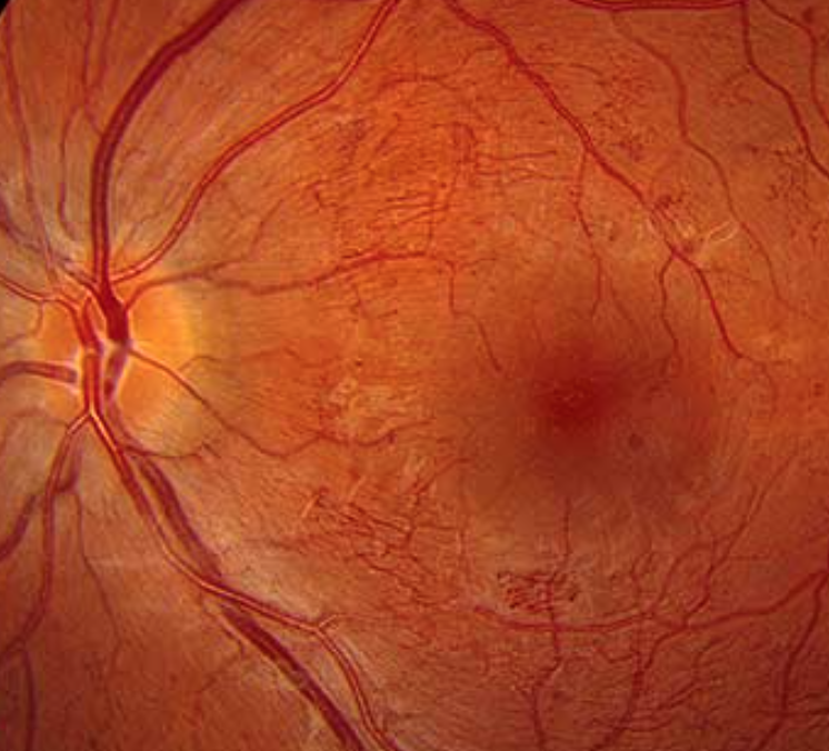
Ophthalmology-Notes And Synopses - Non-proliferative diabetic retinopathy:  intraretinal microvascular abnormality (IRMA; green arrow), venous beading  and segmentation ( blue arrow), cluster haemorrhage (red arrow),  featureless rtina suggestive of non ...
Ophthalmology-Notes And Synopses - Non-proliferative diabetic retinopathy: intraretinal microvascular abnormality (IRMA; green arrow), venous beading and segmentation ( blue arrow), cluster haemorrhage (red arrow), featureless rtina suggestive of non ...

Severe nonproliferative diabetic retinopathy with intra-retinal... |  Download Scientific Diagram
Severe nonproliferative diabetic retinopathy with intra-retinal... | Download Scientific Diagram

Community Eye Health Journal » Recognising and managing diabetic retinopathy

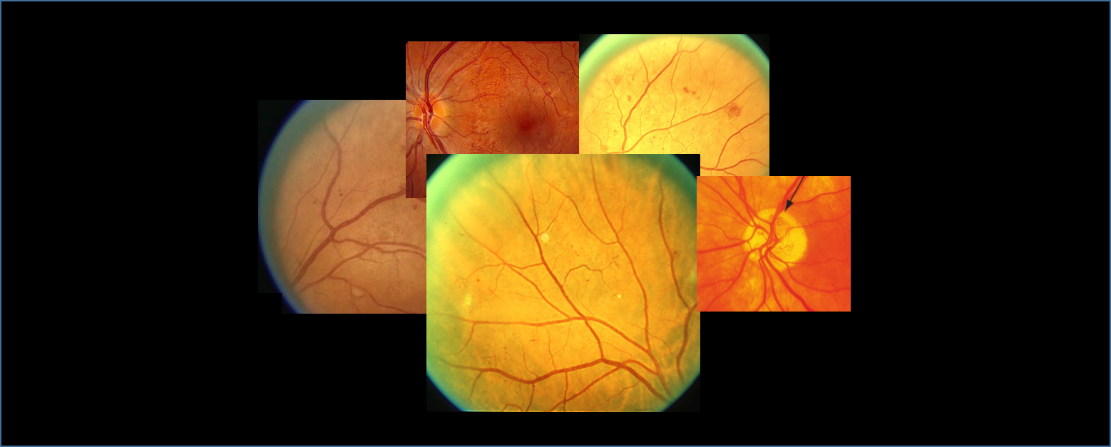
Diabetic Retinopathy – Features of Diabetes : Intraretinal Microvascular  Abnormalities (IRMAs)
Diabetic Retinopathy – Features of Diabetes : Intraretinal Microvascular Abnormalities (IRMAs)

How to diagnose and manage diabetic retinopathy - EyeGuru

OCT in diabetic screening

Diabetic Retinopathy by the Numbers

How to diagnose and manage diabetic retinopathy - EyeGuru

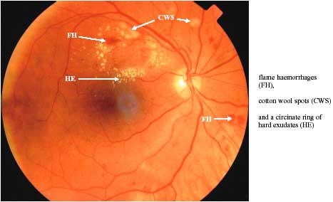
Diabetic Eye Screening Services in Scotland: A Training handbook – July  2003: page 8
Diabetic Eye Screening Services in Scotland: A Training handbook – July 2003: page 8

IRMA vs NVE || intraretinal microvascular abnormality vs neovascularisation  - YouTube
IRMA vs NVE || intraretinal microvascular abnormality vs neovascularisation - YouTube

Diabetic Eye Screening Services in Scotland: A Training handbook – July  2003: page 19
Diabetic Eye Screening Services in Scotland: A Training handbook – July 2003: page 19

GEC | Diabetic Retinopathy ::

Retinal image that exhibits Venous beading and IRMA caused due to... |  Download Scientific Diagram
Retinal image that exhibits Venous beading and IRMA caused due to... | Download Scientific Diagram

Ophthalmology-Notes And Synopses - Intraretinal microvascular abnormalities  (or IRMAs): ▪️IRMAs are shunt vessels and appear as abnormal branching or  dilation of existing blood vessels (capillaries) within the retina that act  to supply
Ophthalmology-Notes And Synopses - Intraretinal microvascular abnormalities (or IRMAs): ▪️IRMAs are shunt vessels and appear as abnormal branching or dilation of existing blood vessels (capillaries) within the retina that act to supply
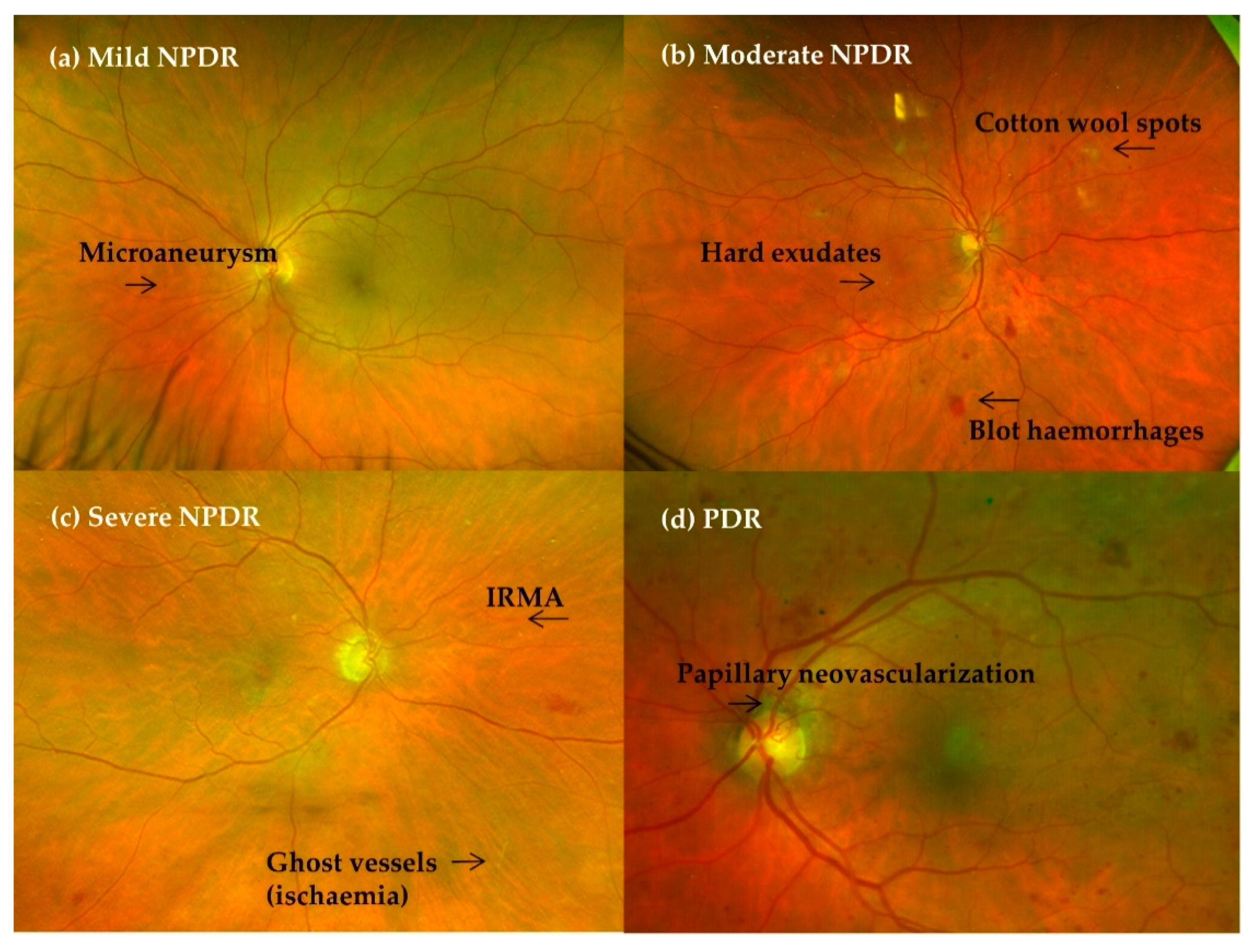
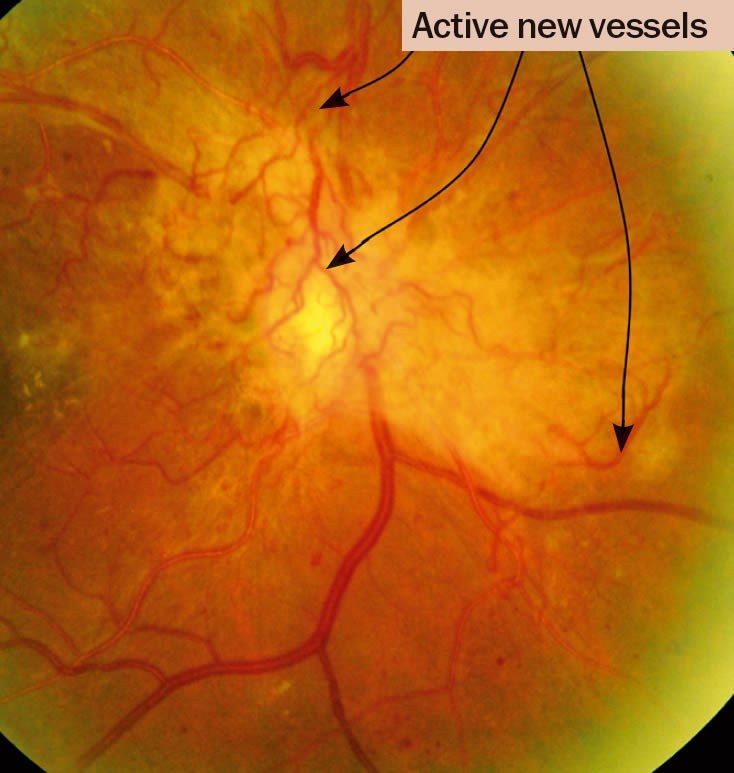
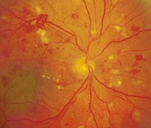
Diabetic retinopathy with intraretinal bleedings, new vessels,... |  Download Scientific Diagram
Diabetic retinopathy with intraretinal bleedings, new vessels,... | Download Scientific Diagram

Diabetic Retinopathy – Features of Diabetes : Intraretinal Microvascular  Abnormalities (IRMAs)
Diabetic Retinopathy – Features of Diabetes : Intraretinal Microvascular Abnormalities (IRMAs)

How to diagnose and manage diabetic retinopathy - EyeGuru

IJMS | Free Full-Text | Role of Oral Antioxidant Supplementation in the  Current Management of Diabetic Retinopathy
IJMS | Free Full-Text | Role of Oral Antioxidant Supplementation in the Current Management of Diabetic Retinopathy
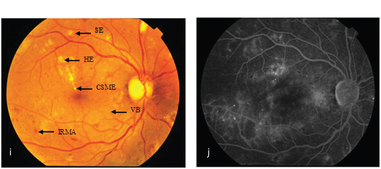
Diabetic retinopathy grading and classification - EyeSteve.com

Diabetic retinopathy grading and classification - EyeSteve.com

Diabetic retinopathy - ScienceDirect

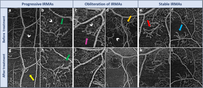
Morphological changes in intraretinal microvascular abnormalities after  anti-VEGF therapy visualized on optical coherence tomography angiography |  Eye and Vision | Full Text
Morphological changes in intraretinal microvascular abnormalities after anti-VEGF therapy visualized on optical coherence tomography angiography | Eye and Vision | Full Text